
airway structure and labels
The airway structure is a crucial component of the respiratory system, facilitating the movement of air in and out of the lungs. Understanding its anatomy is essential for medical professionals and students alike.
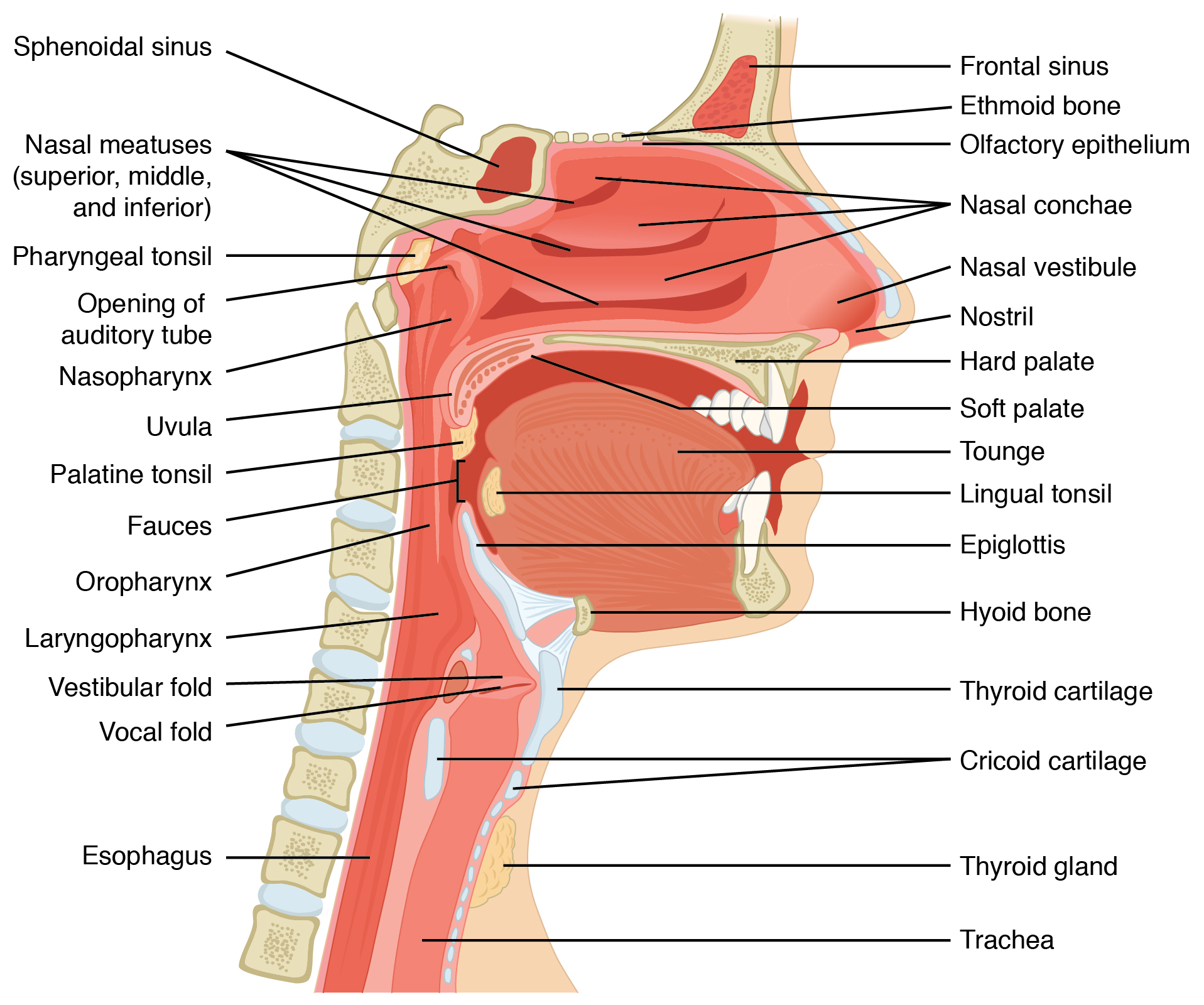
The airway can be divided into two main sections: the upper airway and the lower airway. The upper airway includes the nose, mouth, pharynx, and larynx. Specifically, the pharynx is further segmented into regions such as the nasopharynx (which is located behind the nose) and the oropharynx (located behind the mouth). This anatomy is illustrated in detail in various resources, which provide labeled diagrams to aid in comprehension (MedicTests - Basic Airway Anatomy Guide).
The lower airway consists of the trachea, bronchi, and bronchioles. The trachea branches off into the right and left primary bronchi, which subsequently divide into secondary and tertiary bronchi. This branching continues into smaller bronchioles leading to the alveoli, where gas exchange occurs. Visual diagrams often illustrate these components effectively, making it easier to grasp the overall structure of the respiratory system (Lumen Learning - Organs and Structures of the Respiratory System, Wikipedia - Respiratory Tract).
Additionally, comprehensive resources, such as the Canadian Lung Association (Link - Respiratory System) and Kenhub (Link - Respiratory System Anatomy and Functions), provide in-depth explanations and labeled diagrams that further clarify the various parts of the airway.
For visual representation, a labeled diagram depicting the lungs and respiratory system can be found at SERC (Link - Labeled Diagram of the Lungs/Respiratory System). This collection of resources serves as a valuable reference for understanding airway structure and function.
Sources

Related Questions
Work fast from anywhere
Stay up to date and move work forward with BrutusAI on macOS/iOS/web & android. Download the app today.